A new breast lump deserves assessment even when it is painless, smooth, or mobile, because touch alone cannot identify its cause. You will learn which changes need urgent attention, which tests doctors use, and when monitoring, biopsy, drainage, or breast lump surgery may be appropriate.
Key takeaways
- Seek prompt care for a new, hard, fixed, or rapidly growing lump.
- Do not repeatedly press or massage the lump to monitor it.
- Imaging and tissue diagnosis determine whether biopsy or removal is needed.
- Discuss persistent or changing findings with a breast or general surgeon.
Which breast-lump changes need prompt or emergency medical review?
A hard breast lump or fixed breast lump needs prompt clinical assessment, especially if it is new or persistent. Arrange an appointment rather than repeatedly checking it if it is a rapidly growing breast lump, painless, or accompanied by a new one-sided change.
Skin dimpling is a breast cancer sign that needs assessment, as do ulceration, new nipple inversion, spontaneous or bloody discharge from one nipple, or an enlarged underarm node.
| Urgency | Warning signs | Action |
|---|---|---|
| Same-day or emergency assessment | Red, hot, painful breast with fever, chills, marked swelling, or feeling seriously unwell | Seek urgent care; mastitis or an abscess may need antibiotics, ultrasound-guided drainage, or surgical drainage |
| Prompt outpatient appointment | Persistent or enlarging lump without severe infection symptoms | Arrange clinical examination and imaging; this is not necessarily an emergency |
| Prompt outpatient appointment | Hard or fixed lump, skin dimpling or ulceration, new nipple inversion, one-sided bloody discharge, or enlarged underarm node | Do not wait for the lump to disappear |
Growth alone does not prove cancer, and pain does not reliably separate harmless from serious causes. Most lumps are benign, but touch cannot distinguish a cyst, fibroadenoma, infection, and cancer.
During pregnancy or breastfeeding, infection and benign changes can cause lumps. A lump that persists after mastitis treatment, fails to improve, or remains unexplained still needs imaging and sometimes biopsy. Do not keep delaying review because you are breastfeeding.
How can you monitor a lump without irritating it or guessing its cause?
Use flat finger pads to check a breast lump once, then leave it alone; do not squeeze or massage it repeatedly.
Record the date you first found it, the breast and position using a clock-face and distance from the nipple, approximate size, tenderness, mobility, skin or nipple changes, and whether it varies before or after menstruation.
- A cyst, fibroadenoma, infection or abscess, fat necrosis, and malignant tumour can all feel smooth, firm, or tender. Examination alone cannot reliably distinguish them.
- Most breast lumps are benign, but a smooth, mobile, or painless breast lump still needs assessment. Touch cannot establish the cause.
- During pregnancy or breastfeeding, record changes but do not postpone review. A lump that persists after mastitis treatment, fails to improve, or remains unexplained needs imaging and sometimes biopsy.
| Pain pattern | What it suggests | What to do |
|---|---|---|
| Focal, persistent pain with a palpable abnormality | A local lesion needs explanation | Arrange clinical assessment; do not assume pain excludes cancer |
| Diffuse cyclical breast pain without a mass | Often hormonal breast pain | Track the pattern; it does not usually lead directly to lump-removal surgery |
Growth alone does not prove cancer, but an enlarging or persistent lump deserves prompt review. A normal screening mammogram does not automatically explain it; targeted imaging may still be needed.
Which clinicians and tests determine whether a lump needs tissue diagnosis?
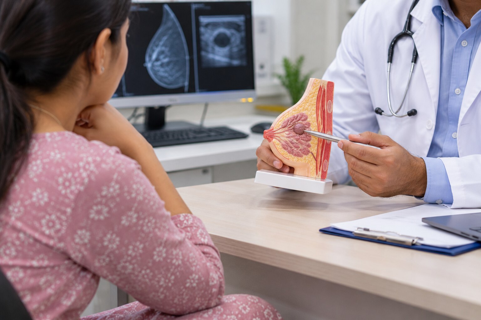
The triple assessment breast lump pathway uses clinical breast examination, breast imaging, and tissue diagnosis when indicated. A GP, breast clinician, or surgeon examines the breast and underarm, then a radiologist selects imaging and compares the result with the examination.
| Age | Usual imaging | Important point |
|---|---|---|
| Under 30 | Breast ultrasound | It is usually the first test for a palpable lump. |
| 30–39 | Ultrasound plus diagnostic mammography or digital breast tomosynthesis when appropriate | The clinician chooses one or both based on examination and risk. |
| 40 and over | Diagnostic mammography or tomosynthesis, often with targeted ultrasound | Targeted imaging evaluates the exact palpable area. |
A normal screening mammogram does not automatically explain a persistent lump. If the examination remains concerning, targeted ultrasound, specialist review, or sampling may still be needed.
For a suspicious solid lesion, an image-guided core-needle biopsy breast lump procedure is commonly preferred. Several tissue cylinders show architecture, help identify tumour type, and allow testing for receptors such as oestrogen receptor, progesterone receptor, and HER2.
Fine-needle aspiration removes cells rather than tissue structure; it can help with selected cysts or lymph nodes, but it is not interchangeable with core biopsy for every solid mass. Family history, previous breast cancer, dense breasts, hormone use, pregnancy, and breastfeeding can alter the imaging sequence or biopsy decision.
The three assessment findings must agree; a benign-looking scan does not override a clinically discordant lump.
When is observation reasonable, and when is biopsy or removal more appropriate?
Finding a lump does not automatically mean breast lump surgery is needed. You may observe a benign breast lump when ultrasound confirms a simple cyst, examination and imaging agree, and symptoms remain manageable. A painful or tense cyst can be aspirated for relief.
| Finding or result | Usual next step | Why it matters |
|---|---|---|
| Simple cyst, matching examination, manageable symptoms | Observation or repeat imaging | No solid component needs treatment |
| Painful or tense cyst | Aspiration | Reduces pressure and discomfort |
| Bloody fluid, residual solid component, or recurrent cyst | Further imaging, core biopsy, or surgical review | These findings need an explanation |
| Suspicious or enlarging solid lesion | Image-guided core-needle biopsy | Provides tissue architecture and tumour information |
| Inconclusive biopsy or disagreement between examination, imaging, and pathology | Specialist or surgical assessment | A benign report does not settle a discordant case |
| Abscess with redness, heat, pain, or fever | Antibiotics plus ultrasound-guided or surgical drainage | This is infection treatment, not elective lump removal |
Breast lump biopsy versus surgery depends on the diagnosis, not preference alone. Removal becomes more appropriate when a lump is enlarging, persistently symptomatic, suspicious on imaging, or unexplained after needle testing. Suspected phyllodes tumour surgery may require complete excision because sampling can miss the lesion’s full pattern.
High-risk or atypical lesions may also need complete removal or additional assessment.
Failure to improve after abscess treatment requires reassessment for a deeper abscess or another cause. A normal screening mammogram does not explain every palpable lump; targeted imaging or tissue diagnosis may still be necessary.
When should you discuss the findings with a breast or general surgeon?
Arrange a breast surgeon consultation in Pune when the next decision is unclear, not only when surgery has been advised. A surgeon can review a growing or symptomatic lump alongside the examination, imaging, and pathology report, then explain whether observation, repeat imaging, biopsy, drainage, or removal fits the evidence.
Discuss a breast lump surgical evaluation when:
- The imaging and needle biopsy do not agree, or the pathology report is inconclusive.
- The lump remains unexplained, enlarges, causes focal persistent pain, or continues after presumed mastitis has been treated.
- Removal is being considered for symptoms, a high-risk or atypical lesion, or a suspected phyllodes tumour.
Excisional surgery is not automatic. Diffuse cyclical breast pain without a palpable mass does not usually lead directly to lump removal, while a persistent mass needs assessment even when it is painless.
Dr Bharat Oza - Laparoscopic Surgeon can be one clinician to discuss breast lump findings and possible surgical options in Pune; the appropriate next step still depends on the examination, imaging, and pathology.
For a breast lump in men, a tender, rubbery lump directly beneath the nipple often reflects gynecomastia. A hard eccentric mass, nipple retraction or discharge, skin change, or enlarged armpit node needs assessment because gynecomastia versus breast cancer cannot be settled by feel alone.
Bring prior reports and images, medication and hormone-use details, family history, and a timeline of changes.
Related service
Breast Lump Surgery Specialist in Wakad, PCMC, Pune Breast lump surgery, also known as lumpectomy or wide local excision, is a procedure to remove a lump (tumor) from the breast, along with a small... View service → |
Frequently asked questions
Which breast-lump changes need prompt or emergency medical review?
Arrange prompt medical assessment for a new, hard, fixed, persistent, painless, or rapidly growing lump. Seek urgent care for a lump with severe redness, fever, rapidly worsening swelling, skin breakdown, or feeling acutely unwell.
How can you monitor a breast lump without irritating it?
Note when you found it, its location, approximate size, tenderness, and changes in the skin or nipple. Check it gently at set intervals instead of repeatedly pressing, squeezing, or massaging it.
Which clinicians and tests determine whether a breast lump needs tissue diagnosis?
A primary-care doctor, breast physician, radiologist, or surgeon can assess the lump. Evaluation may include a clinical examination, diagnostic mammogram, targeted breast ultrasound, and image-guided core needle biopsy when imaging or examination requires tissue confirmation.
When is observation reasonable, and when is biopsy or removal more appropriate?
Observation can be reasonable when examination and imaging show a clearly benign finding and a clinician gives you a follow-up plan. Biopsy is appropriate when the lump is suspicious, persistent, enlarging, or imaging is unclear; removal may be discussed for confirmed or strongly suspected disease, symptoms, growth, or diagnostic uncertainty.
When should you discuss breast-lump findings with a surgeon?
Discuss the findings with a breast or general surgeon when a lump persists, grows, feels fixed, produces concerning imaging, needs biopsy or excision, or remains unexplained after initial assessment.