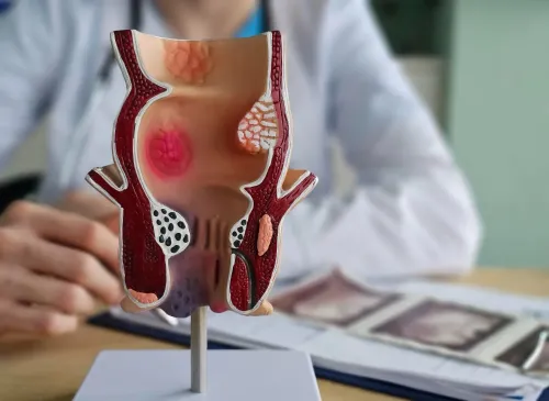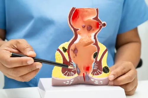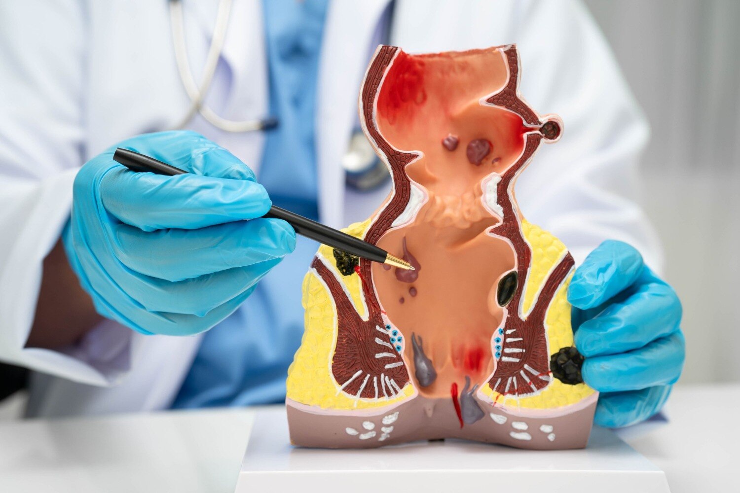
A small opening that repeatedly leaks pus, blood-stained fluid or foul-smelling discharge deserves a closer look, especially after a painful swelling has drained. By the end, you will know which symptoms suggest an abscess, what clinicians examine, when imaging helps and how tract anatomy influences fistula treatment.
Key takeaways
- Repeated pus or blood-stained discharge from one opening needs medical assessment.
- Seek urgent care for fever, rapidly worsening pain, or spreading redness.
- MRI or endoanal ultrasound maps fistula tracts hidden beneath the skin.
- Treatment depends on whether the tract crosses the anal sphincter muscles.
How can you tell fistula drainage from other causes?
Fistula drainage is more concerning than ordinary mucus when a small external opening near the anus repeatedly releases pus near anus or blood-stained discharge, especially with a foul odour.
A small, fixed opening that refills after it drains points towards a persistent tract. Pain that eases after fluid escapes is another clue, although it does not confirm a fistula.
- An abscess usually causes a tender, tense swelling with worsening pain. It is less likely to present as a stable opening that repeatedly drains.
- An infected skin cyst sits within the skin and may have a visible central pore. Its swelling is usually localised to the cyst itself.
- Hidradenitis suppurativa tends to produce multiple painful nodules or draining sites in the groin, buttocks or armpits rather than one consistent opening.
- Haemorrhoids more often cause bright-red bleeding, itching or a lump associated with bowel movements. Repeated pus is not typical.
- Normal mucus is usually small in amount and lacks a fixed opening, foul odour, pus or recurrent swelling.
An opening is not proof of a fistula. Pilonidal disease, Crohn’s disease, a sexually transmitted infection and other skin conditions can look similar. Direct perianal examination, and sometimes a gentle rectal examination, is needed to identify the actual cause rather than relying on the drainage pattern alone.
Which swelling needs urgent assessment rather than routine planning?
An anal abscess needs prompt medical assessment; a fistula usually needs planned evaluation after acute infection is controlled. An abscess is a collection of infected fluid, while a fistula is a persistent tunnel that commonly remains after an abscess drains.
| Abscess | Fistula |
|---|---|
| Tender, tense swelling with worsening pressure or pain | Small opening with repeated pus or blood-stained discharge |
| May cause fever and chills or spreading redness | May settle temporarily after drainage, then recur |
| Requires appropriate drainage | Requires assessment to map the tract and internal opening |
Seek urgent medical care if you have:
- Rapidly increasing pain or swelling
- Spreading redness into nearby skin
- Fever and chills
- Difficulty sitting
- Inability to pass stool or urine
- A sudden feeling of being acutely unwell
These signs can indicate an anal abscess or spreading infection. Do not wait for an elective fistula appointment.
Symptoms can return after an abscess drains because the internal opening and inflamed tract remain after pressure falls. The external opening can seal, allowing another painful collection to form. Repeated squeezing, puncturing or self-drainage does not prove healing; it can injure skin or delay proper drainage.
Antibiotics alone do not usually remove a typical cryptoglandular fistula. They may accompany treatment for cellulitis, systemic infection or immunosuppression, but an abscess still requires appropriate drainage.
What happens during diagnosis, and when is imaging useful?
Before the appointment, write down the pattern rather than relying on memory. Note when the first episode began, how many times swelling or discharge returned, the exact location, the amount and colour of fluid, its odour, fever, bowel changes, previous drainage or surgery, Crohn’s disease, diabetes, and medicines affecting immunity or bleeding.
1. A fistula examination usually starts with inspection of the perianal skin. The clinician looks for an external opening, redness, discharge, tenderness, induration and an active abscess.
2. When appropriate, the clinician performs a gentle digital rectal examination to assess pain, swelling and possible internal openings. Do not assume the visible opening shows the entire tract; branches or the internal opening can lie deeper.
3. Pelvic MRI is particularly useful for recurrent, complex, horseshoe or Crohn-related fistulas. It can map branches, hidden abscesses and the relationship with the sphincter before treatment.
4. Endoanal ultrasound helps in selected cases by assessing the tract and sphincter. Ask whether it will answer a specific question that MRI cannot.
5. Examination under anaesthesia is useful when pain prevents a clear examination or the tract remains uncertain. It allows the surgeon to map the tract, identify an internal opening and find secondary extensions.
A scan does not replace clinical assessment, and a draining opening does not prove a fistula. Hidradenitis suppurativa, an infected cyst, pilonidal disease, Crohn’s disease and sexually transmitted infections can look similar, so examination findings determine the next step.
Why does the tract’s anatomy change the treatment?
The decisive question is how much of the external anal sphincter the tract crosses, not how small the skin opening looks. That measurement separates a simple versus complex fistula and determines the balance between reliable drainage, healing time and continence risk.
A low, simple tract with limited anal sphincter involvement may suit fistulotomy: the surgeon opens the tract so it heals from its base. Dividing too much muscle can impair continence.
| Option | What it does | Main trade off or use |
|---|---|---|
| Fistulotomy | Opens the tract along its length | Effective for selected low tracts, but recurrence, delayed healing and continence problems remain possible |
| Loose seton placement | Passes a thread through the tract to keep drainage flowing | Controls sepsis before definitive treatment; it is not automatically curative |
| LIFT | Closes the tract between the sphincter muscles | Preserves sphincter muscle, but healing and recurrence vary |
| Endoanal advancement flap | Covers the internal opening with tissue | Avoids deliberate sphincter division, though failure and recurrence can occur |
| Laser or other minimally invasive closure | Treats the tract through a limited approach | Not interchangeable with standard operations or universally superior |
Higher, branched, recurrent or Crohn related fistulas often need staged or sphincter preserving treatment rather than immediate fistulotomy. Discuss recurrence, delayed healing and continence before choosing an operation. Outcomes depend on the tract’s anatomy, previous surgery and follow up, so laser closure is not a guaranteed shortcut.
When should you arrange specialist evaluation?
Arrange a recurrent fistula evaluation when a small opening repeatedly drains, swelling returns after bursting, or previous treatment has not produced lasting healing.
1. Seek further investigation for an unusually hard or enlarging area, unexplained bleeding, weight loss, or a wound that fails to heal. These findings do not prove cancer, but do not dismiss them as an ordinary abscess; examination or tissue sampling may be needed.
2. Tell the clinician if you have Crohn’s disease. A Crohn’s-related fistula requires assessment for active rectal or perianal inflammation and coordinated medical treatment. Aggressive fistulotomy can be unsuitable when inflammation is substantial or the tract crosses important sphincter muscle.
- Where is the internal opening?
- Do I need pelvic MRI or examination under anaesthesia?
- How much of the external anal sphincter does the tract cross?
- Will treatment be staged?
- How will delayed healing, continence and recurrence be monitored?
4. Do not choose treatment from the skin opening alone. A tiny opening can connect to a high, branched or horseshoe tract, while MRI or examination under anaesthesia can reveal extensions that change the safest approach.
In Pune, Dr Bharat Oza - Laparoscopic Surgeon can assess these anatomy-based decisions, including whether a sphincter-preserving plan is safer than fistulotomy. Share prior drainage, surgery, Crohn’s disease, diabetes, and medicines affecting immunity or bleeding.
Related service
Fistula Treatment Specialist in Wakad, PCMC What is Anal Fistula? An anal fistula is a tunnel connecting the skin near the anus to the inside of the bowel (usually the rectum). View service → |
Frequently asked questions
How can you tell fistula drainage from other causes?
Fistula drainage usually comes repeatedly from a small opening near the anus and contains pus or blood-stained fluid, often with a foul odour. Mucus alone does not prove a fistula, so a clinician must examine the area.
Which swelling needs urgent assessment rather than routine planning?
Seek urgent assessment for rapidly increasing swelling, severe or throbbing pain, fever, chills, spreading redness, or difficulty passing urine. These signs can indicate an abscess requiring prompt treatment.
What happens during diagnosis, and when is imaging useful?
A specialist checks the skin opening, tenderness, drainage and anal canal, sometimes using anoscopy. MRI of the pelvis or endoanal ultrasound helps locate branches, abscesses and deep tracts that examination cannot show.
Why does the tract’s anatomy change the treatment?
The surgeon must know how much of the anal sphincter the tract crosses. A superficial tract may allow fistulotomy, while a deeper or branching tract may need staged or sphincter-sparing treatment.
When should you arrange specialist evaluation?
Arrange evaluation when discharge recurs, the opening closes and swelling returns, or an abscess has drained but does not heal. Prompt review is especially important after repeated episodes or previous anal surgery.