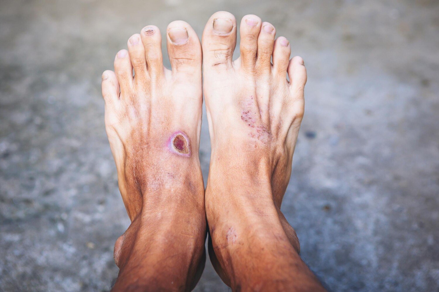
A diabetic foot wound that stays open is not automatically a reason for amputation, but it is a reason to identify why healing has stalled. By the end, you will know which changes require hospital care, how to track the wound, what tests guide treatment, and when procedures such as debridement, drainage, revascularisation, or amputation enter the discussion.
Key takeaways
- Seek reassessment when the wound heals less than 50% after four weeks.
- Keep pressure off the wound and check it daily for worsening changes.
- Ask about blood-flow tests, infection imaging, and tissue sampling when indicated.
- Discuss debridement, drainage, revascularisation, or amputation based on the cause.
How long can a diabetic foot wound stay open before it needs surgical assessment?
A non-healing diabetic foot wound needs reassessment when it shows no substantial improvement after about four weeks of good wound care, infection treatment, pressure relief, and blood-glucose management. IWGDF guidance treats healing of less than 50% within four weeks as a reason to investigate peripheral arterial disease and consider vascular imaging or revascularisation.
Do not wait four weeks if the wound is worsening. Seek urgent hospital assessment for:
1. Spreading redness or swelling, warmth, pus, foul odour, fever, confusion, or rapidly increasing wound size or discoloration. These signs can indicate severe infection, an abscess, gangrene, or a necrotising infection.
2. Black tissue, exposed tendon or bone, or a deep wound with new swelling. A black tissue diabetic foot ulcer can reflect gangrene or tissue death, while exposed bone raises concern for deep infection.
3. A cold or pale foot, severe pain, or sudden swelling and tightness. These findings can signal severe ischemia or compartment syndrome and may require urgent diabetic foot surgery.
Neuropathy can remove pain, so a painless ulcer can still be dangerous. A small, clean, stable wound without these warning signs can receive planned outpatient evaluation, but routine scheduling is unsafe when infection or blood-flow loss is progressing.
Prompt review does not automatically mean amputation; it identifies whether drainage, debridement, revascularisation, or another treatment is needed.
How can you monitor the wound and protect it while waiting for review?
Clean the wound exactly as instructed, cover it with the recommended dressing, and keep weight off it until review. For a neuropathic plantar forefoot or midfoot ulcer, a non-removable knee-high offloading device is generally the first choice when safe and tolerated.
Specialised footwear or another offloading device is safer than continuing to walk on the ulcer.
1. At every dressing change, record a diabetic foot wound measurement: longest length, widest width, and deepest point in millimetres or centimetres. Use the same method each time; an apparently smaller measurement taken differently can mislead you.
2. Use wound photography to track change. Photograph the wound from the same distance, place a ruler beside it, and record the date. Note redness, warmth, swelling, callus, skin colour, drainage amount and colour, and any new odour.
3. Check both feet daily. Neuropathy and painless foot ulcer complications can progress without the pain that would normally prompt action. Seek urgent medical assessment for spreading redness or swelling, warmth, pus, foul odour, fever, confusion, rapidly increasing size or discoloration, black tissue, or a cold or pale foot.
4. Do not cut callus, probe deeply, apply household antiseptics, or soak the foot unless a clinician tells you to. Sharp removal of callus or dead tissue is standard care, but limited clinic debridement differs from an operating-room procedure for extensive necrosis, infection, abscess, or deeper tissue involvement.
What tests show whether the wound needs surgery?
Clinicians decide whether surgery is needed by combining diabetic foot ulcer assessment, infection testing, imaging, and circulation measurements. A non-healing wound alone does not mean amputation; the cause determines the treatment.
1. Examine depth, drainage, odour, surrounding redness or swelling, foot temperature, pulses, sensation, deformity, and whether a sterile probe reaches bone. A deep wound, exposed bone, or a positive probe finding raises concern for osteomyelitis or a deep abscess.
2. Obtain a foot X-ray to look for gas, a foreign body, bone destruction, or deformity. MRI, or another scan, helps when osteomyelitis or a deep abscess remains uncertain.
3. Use blood tests to assess inflammation, infection, kidney function, and glucose control. A surface swab does not prove that antibiotics or surgery are needed because it can grow colonising bacteria. When infection is suspected, cleaning and debridement followed by a deep tissue culture gives more useful results.
A bone culture is preferred when osteomyelitis is suspected and bone sampling is feasible.
4. Check blood flow with pulses, Doppler waveforms, toe pressure, or skin-perfusion measurements. An ankle-brachial index in diabetes can appear normal despite ischemia because calcified arteries resist compression. If circulation is reduced, duplex ultrasound, CT angiography, MR angiography, or catheter angiography can map blockages before revascularisation or tissue removal.
Which diabetic foot surgery might be considered, and when?
The procedure must match why the wound is not healing. A painless ulcer does not automatically need amputation; infection, trapped pus, poor blood flow, pressure, dead tissue, deformity, and bone involvement require different solutions.
| Procedure | What it addresses | When it may apply |
|---|---|---|
| Diabetic foot debridement | Dead or infected tissue, callus, foreign material | Limited removal in a clinic; operating-room treatment when necrosis, deep infection, tendon or joint involvement, or extensive tissue loss is present |
| Abscess drainage | Pus trapped in a deep space | Drainage is essential; antibiotics alone cannot replace it |
| Diabetic foot osteomyelitis surgery | Infected or necrotic bone | When infection is extensive, uncontrolled with medical treatment, or preventing closure; selected forefoot cases may respond to antibiotics alone if there is no urgent drainage need, uncontrolled soft-tissue infection, or severe peripheral arterial disease |
| Corrective surgery | Repeated pressure from a fixed deformity | Tendon balancing, metatarsal-head or bony-prominence surgery, or selected Charcot reconstruction after infection and circulation problems are controlled |
| Revascularisation diabetic foot ulcer treatment | Arteries unable to supply healing tissue | Restore blood flow before tissue removal when ischemia threatens healing |
| Skin grafting or amputation | Coverage or removal of non-viable tissue | Grafting covers a prepared wound; amputation preserves the limb when viable tissue cannot be saved |
Without bone resection, osteomyelitis treatment lasts about six weeks. After minor amputation with a positive bone margin, up to three weeks may be considered. A consultation with Dr Bharat Oza - Laparoscopic Surgeon can help match the proposed operation to examination, imaging, circulation, infection control, and closure prospects.
Related service
Best Diabetic Foot Surgery Specialist in Wakad, PCMC, Pune Diabetic foot surgery involves a range of procedures to treat complications like ulcers, infections, deformities, and poor circulation in individuals... View service → |
What should you ask before agreeing to a diabetic foot operation?
A diabetic foot surgery consultation should begin with one question: why has the wound failed to heal? Ask the team to identify infection, poor circulation, pressure, deformity, exposed or dead tissue, or bone involvement. A non-healing wound alone does not settle an amputation decision in diabetic foot disease.
| Option | What it means | Ask when |
|---|---|---|
| Clinic debridement | Removal of callus or dead tissue | Necrosis is limited and infection is controlled |
| Hospital drainage | Opening and draining trapped pus | A deep abscess or spreading infection is present |
| Bone removal | Removing infected or non-viable bone | Osteomyelitis prevents control or closure |
| Deformity correction | Reducing repeated pressure | Offloading failed after infection and ischemia are addressed |
| Revascularisation | Restoring arterial blood flow | Testing shows inadequate flow for healing |
| Grafting or amputation | Covering a prepared wound or removing unsalvageable tissue | Viability and closure prospects require it |
Ask these questions before consenting:
- What finding would change this plan?
- Will you collect a deep-tissue or bone sample after cleaning, rather than relying on a surface swab?
- How was blood flow measured: pulses, Doppler, toe pressure, skin-perfusion testing, or imaging?
- How will glucose control and kidney function affect contrast scans, antibiotics, anaesthesia, or healing?
- What device will provide postoperative offloading, and for how long?
- How long will antibiotics continue? Which fever, spreading redness, drainage, worsening swelling, confusion, or colour change requires immediate hospital return?
A multidisciplinary diabetic foot team may include a surgeon, diabetologist, infectious-disease clinician, vascular specialist, podiatry or wound-care professional, and rehabilitation staff. Do not remove only visible black tissue or keep walking on the wound; confirm that deeper infection and ischemia have been addressed.
Frequently asked questions
How long can a diabetic foot wound stay open before surgical assessment?
Arrange reassessment when the wound shows no substantial improvement after about four weeks of good care. Healing of less than 50% in four weeks warrants investigation for peripheral arterial disease and possible vascular imaging or revascularisation.
How can you monitor and protect a diabetic foot wound while waiting for review?
Inspect the wound and surrounding skin daily, keep the dressing clean and dry, follow pressure-relief instructions, and monitor for increasing redness, swelling, drainage, odour, pain, fever, or blackened tissue. Seek urgent care for rapidly worsening infection or reduced blood flow.
What tests show whether a diabetic foot wound needs surgery?
Assessment can include wound measurement, pedal-pulse examination, ankle-brachial index, toe pressure, Doppler ultrasound, foot X-ray, MRI for suspected bone infection, and blood tests. Clinicians may also culture properly collected tissue when infection requires targeted antibiotics.
Which diabetic foot surgery might be considered, and when?
Procedures depend on the cause: debridement removes dead tissue, drainage treats an abscess, revascularisation improves blood flow, and limited amputation removes non-viable or infected tissue when preservation is not possible. Treating pressure or deformity can help prevent recurrence.
What should you ask before agreeing to a diabetic foot operation?
Ask what the operation will fix, what alternatives exist, how much tissue may be removed, whether blood flow must be restored first, how infection will be treated, the expected recovery and off-loading plan, and how recurrence risk will be reduced.